At the St George Peritonectomy & Liver Cancer Unit we provide the following radiological studies:
CT scan
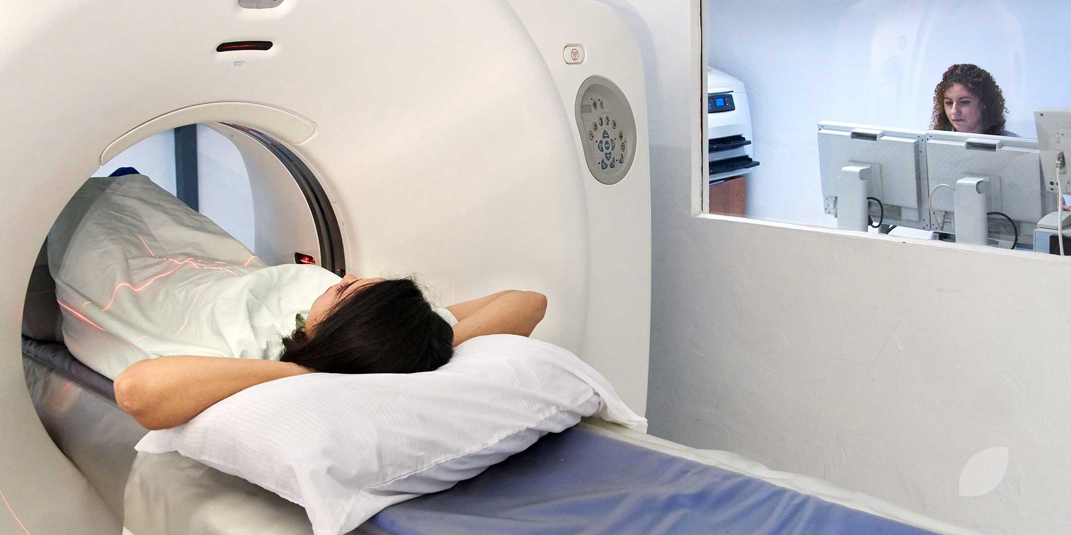
A CT (computerised tomography) scan uses x-ray beams to create a detailed picture of the inside of the body
- Before the scan, you may be given a special drink and a dye injected into a vein to make the pictures This may make you feel hot all over for a few minutes and leave a strange taste in your mouth.
- The CT scanner is large and round like a You will lie on a table that moves in and out of the scanner.
- It takes about 30 minutes to set up the machine, and the scan itself takes 5–10.
MRI scan (with or without Primovist contrast)
An MRI (magnetic resonance imaging) scan uses radio waves and magnetism to create cross-sectional pictures of the body. This is likely to incur a cost at your expense. Please discuss if there are financial issues and we will arrange an alternate test (angiogram, details below).
- Dye may be injected into a vein before the scan to help make the pictures clearer.
- You will lie on a table that slides into a metal cylinder that is open at both ends.
- Some people feel anxious lying in the narrow metal cylinder. You may be given a mild sedative to help you relax.
- Before arranging the test, your doctor will ask you questions about your medical history to check you can have the test. People who have a pacemaker or other metallic objects in their body cannot have an MRI.
- The MRI machine can be noisy, you may be offered ear protection.
PET scan
During a PET (positron emission tomography) scan you will be injected with a small amount of radioactive glucose solution.
- It takes 30–90 minutes for the solution to circulate around your You will be asked to sit quietly during this time.
- Your body is then scanned for high levels of radioactive glucose.
- Cancer cells show up brighter on the scan because they are more active and take up more of the glucose solution than normal cells.
- It may take several hours to prepare for and have a PET scan, it is usually done on an outpatient basis.
Angiogram Procedures
This is a two-part procedure performed at St George Hospital. During the first phase, a catheter will be inserted into an artery in your groin under local anaesthetic, this is used to introduce dye into your liver to highlight any possible disease. The second phase involves a CT scan to scan the liver while the dye is being injected. Once the angiogram has been completed, the catheter will be removed and radiology staff will observe the puncture site to ensure that bleeding does not occur.
Depending on the size of the catheter used in your artery, you will be required to stay under observation in the Radiology Department for a minimum of 3 to 4 hours.
- This is not a fasting test, a light breakfast is permitted morning of
- You must be accompanied and driven home by a responsible adult or you may go home alone in a taxi with a responsible adult waiting at the other end. A friend or relative must stay with you
- You must not drive nor do any heavy lifting (eg. shopping or vacuuming) or excessive walking for 24 hours after the
- Please do not shower until the day after your
- We ask that you inspect the insertion site daily for approximately 1 week. You may see a small bruise or feel a small lump. This is normal.
- However, should any swelling or bruising NEWLY appear or INCREASE in size, this may mean that your artery may be bleeding. If either occurs, lie flat and either yourself or a friend apply and maintain firm pressure over the entry site. Call 000 for an ambulance to take you to the nearest
If you have any questions/ queries regarding the angiogram procedure please contact the St George Hospital Radiology Nurse Unit Manager / In Charge RN on (02) 9113 3664 from 7.30am and 9pm. Alternatively, contact the St George Hospital Emergency Department (out of hours) on (02) 9113 1680.